|
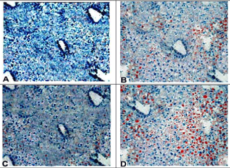
| Figure 6: Photomicrographs of liver specimens showing more quantitatively and qualitatively extensive parenchymal fatty infiltration (prominent red fat globules) in group IV rat (D) as compared to group II (B). Liver specimen of the group III animal (C) showed mild or minimal fatty infiltration while that of control rat (A) was devoid of the fatty infiltration (Oil red stain x100). |