|
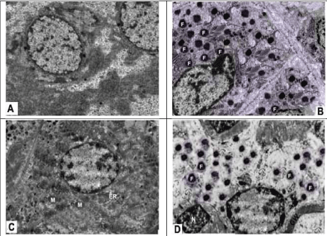
| Figure 9: Electromicrographs of hepatic and renal specimens of group I and group IV animals. There were extensive black fat globules (F) indented the nucleus of hepatocyte (B) as well as extensive black fat globules (F) detected above the nucleus (N) of renal tubular cells with thickening of the glomerular wall (GW) caused by diabetes mellitus (D). In contrast, no fatty globules were detected within the hepatocytes (A) nor within the renal tubular cells (C) that showed normal mitochondria (M) and endoplasmic reticulum (ER) (Transmission electron microscope × 10,000). |