|
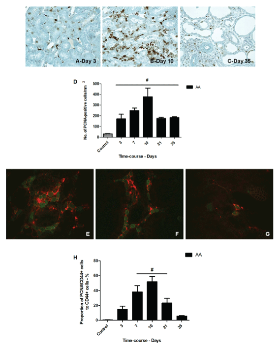
| Figure 5: Representative photomicrographs of PCNA-positive cells in the tubules and the interstitium (x400) in AA-treated rats at day 3 (A), day 10 (B) and day 35 (C). Semiquantitative analysis of PCNA-positive staining in AA-treated rats at different time3 points after treatment (D). Representative photomicrographs of double immunostaining (x1000, arrow) for CD44 and PCNA in AA-treated rats at day 10 (E-G). As shown, CD44 (red) was localized on the basolateral and apical cellular membrane while PCNA labeled the cellular nuclei (green). Semi-quantitative analysis of CD44/PCNA-positive staining in tubular and interstitial cells in control or AAtreated rats at different time points after treatment (H). #p ≤ 0.05 versus control |