|
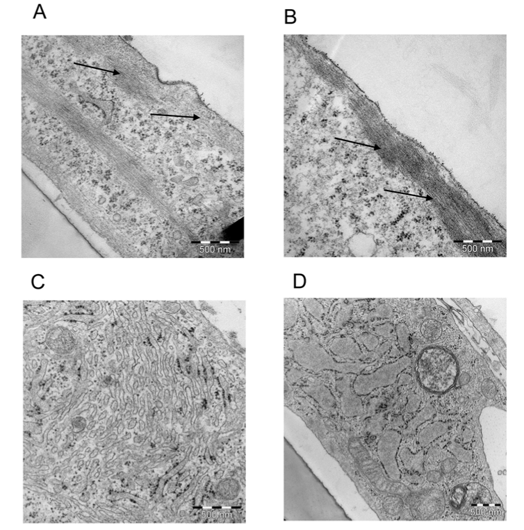
| Figure 6: Representative transmission electron micrograph of SSEA-1+ cells after differentiation protocol. (A and B) Differentiated SSEA-1+ cells revealed large areas of actin organized along the cell membrane as functional unit (arrows). (C and D) SSEA-1+ cells in growth medium. |