|
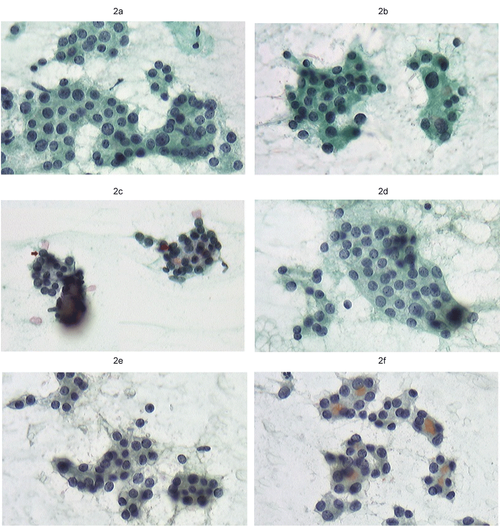
| Figure 2: Histology: Indeterminate cell images of A. nodule. a: Discriminant analysis: A. nodule small, nonspherical nuclei no chromatin condensation. b: Discriminant analysis: Papillary ca indistinguishable cell image with irregular nuclei, light and shade, and nuclear grove. c: Discriminant analysis: A. nodule nuclear pseudo-inclusion small, unclear intranuclear inclusion, not suggesting Papillary ca. d: Discriminant analysis: A. nodule nuclear groove flat nuclei with weak staining and mild atypia. e: Discriminant analysis: Papillary ca intranuclear inclusion with partially unclear boundary Indistinguishable aggregates whose three-dimensional nuclear structure and 3D-CV are similar to those of Papillary ca. f: Discriminant analysis: Papillary ca nuclear grove Indistinguishable aggregates whose three-dimensional nuclear structure and 3D-CV are similar to those of Papillary ca. |