|
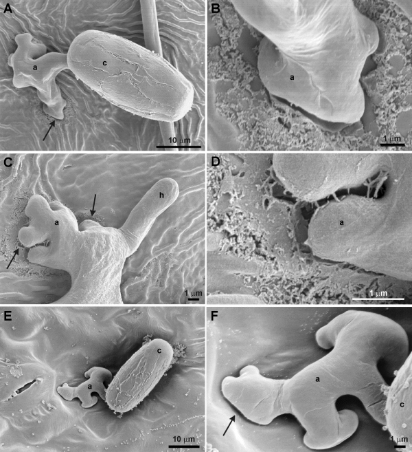
| Figure 2: Degradation of the cuticular layer of Vitis vinifera cv. Chasselas upon contact with infectious structures of Erysiphe necator observed by scanning electron microscopy. (A) Appressorium formation from a conidium and the first step of cuticular degradation (arrow). (B) Detail of the zone of contact of the appressorium with the cuticular layer, which is partially depolymerised. (C) Functional appressorium displaying active degradation of the cuticular layer (arrows) and the developing primary hypha. (D) Detail of C showing an important erosion of the cuticular surface. (E) Leaves pre-treated with the inhibitor DIPF showing no cuticular degradation neither under the conidium nor the appressorium. (F) Detail of (E) showing no erosion of the cuticular surface (arrow). a: appressorium, c: conidium, and h: hypha. |