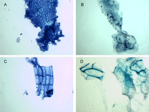
A: Brussels sprouts, MGG stain, 40x magnification
B: Brussels sprouts, PAP stain, 60x magnification
C: Carrot, MGG stain, 40x magnification
D: Carrot, PAP stain, 60x magnification
|
| Figure 4: Brussels sprouts on the MGG stain show compactly arranged sheets of cells that are dense and polygonal in shape, resembling fish scale (A).
Their cell walls are moderately thick. The Pap stain shows a nest of overlapping, plump round cells, which appears to bloom from a stalk-like portion (B).
Compared to the rigid assembly of the cells in (A), these cells are delicate and translucent. The MGG smear of carrots shows rectangular cells stacked endto-
end, forming several orderly arranged tube-like column (C). The Pap stained image reveals a prismatic structure with polygonal-shaped surface and thick
refractive edges (D). A: Brussels sprouts, MGG stain, 40x magnification B: Brussels sprouts, PAP stain, 60x magnification C: Carrot, MGG stain, 40x magnification D: Carrot, PAP stain, 60x magnification |