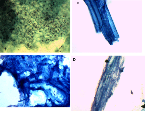
A: Cauliflower, PAP stain, 60x magnification
B: Celery, MGG stain, 40x magnification
C: Cucumber, MGG stain, 60x magnification
D: Cucumber, PAP stain, 100x oil magnification
|
| Figure 5: Pap smear of cauliflower reveals a tightly packed sheet of varied polygonal-shaped cells, each clearly separated by a prominent cell wall (A). At a
low power, they resemble squamous cells in sheets. A microscopic feature of celery cells include long, narrow cylindrical and spiral tubes arranged side by
side (B). The continuous tube contains elliptical pits distributed throughout its length and resembles a nematode larva.
Smears of cucumber demonstrate a formless sheath encasing an elongated structure with ovoid shaped rings (C). The rings appear to form loose curvy spirals
with central line in some section which can mimic a nematode. The Pap image shows a dark woven pattern that appears to be etched onto tissue fragment (D). A: Cauliflower, PAP stain, 60x magnification B: Celery, MGG stain, 40x magnification C: Cucumber, MGG stain, 60x magnification D: Cucumber, PAP stain, 100x oil magnification |