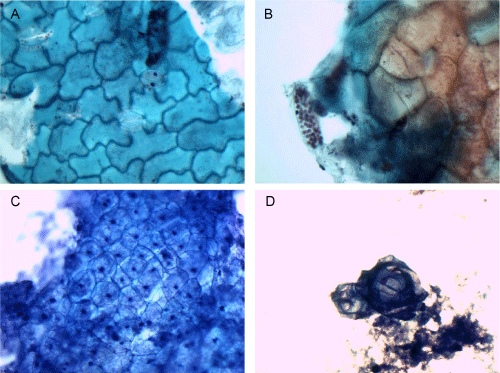
A: Cilantro, PAP stain, 60x magnification
B: Cilantro, PAP stain, 60x magnification
C: Corn, MGG stain, 60x magnification
D: Corn, MGG stain, 60x magnification
|
| Figure 6: Cells of the cilantro have a wavy shaped outline and are closely linked in a jigsaw-like pattern. The thick cell wall appears rigid, and the cytoplasm
appears mostly dense and homogenous with light flecks of reddish, pigmented granules. Sprouting from the tissue is a tubular structure packed with numerous,
rounded beads (A). Notably, stomata are interspersed among the tissue fragments (B). The stomata appear as slit-like pores flanked by two reniform-shaped
guard cells. Kernels of corn have plump polygonal cells with rounded edges on MGG stain (C,D). The fine cell wall surrounds the cell that has a foamy but
granular cytoplasm. Centrally in each cell, there is a small, round nuclei. Thin balloon-shaped cells appear in a loose arrangement and resemble squamous
cells in sheets and anucleated squamous cells singly (D). A: Cilantro, PAP stain, 60x magnification B: Cilantro, PAP stain, 60x magnification C: Corn, MGG stain, 60x magnification D: Corn, MGG stain, 60x magnification |