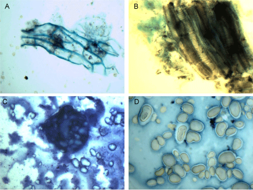
A: Eggplant seeds, PAP stain, 60x magnification
B: Eggplant, PAP stain, 60x magnification
C: Fava beans, MGG stain, 60x magnification
D: Fava beans, PAP stain, 60x magnification
 |
| Figure 7: Eggplants display a diverse, multicolored morphology. The PAP smear of eggplant flesh shows several conducting tubes are bundled together with
regularly spaced elliptical pits traversing through it (A). PAP smear of eggplant seeds contains several rows of oblong, quadrilateral cells, with thick refractile
blue cell walls (B). Fava beans are smeared into bean-shaped fragments that are singly dispersed or loosely aggregated (D) with a thick cell wall that does not
take up PAP staining. The MGG stain shows a dark staining, purple wall (C). A: Eggplant seeds, PAP stain, 60x magnification B: Eggplant, PAP stain, 60x magnification C: Fava beans, MGG stain, 60x magnification D: Fava beans, PAP stain, 60x magnification |