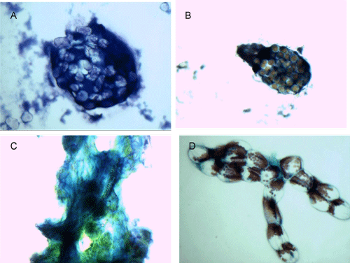
A: Green beans, MGG stain, 60x magnification
B: Green beans, PAP stain, 40x magnification
C: Lettuce, MGG stain, 60x magnification
D: Lettuce, PAP stain, 60x magnification
|
| Figure 9: Green beans consist of small round cells packed together in aggregated clusters (A, B). In the Pap stain, the cell wall stains more prominently as
a light blue color, and the cytoplasm takes up an orange color (B). Almost all green bean cells are clustered as shown, and resemble conidiophores from
Coccidiomycosis. Leaves of lettuce (C, D) have display numerous papillary fronds branching from a thick stalk with helical spiral tubes on MGG stain (C). Many
large, red-pigmented granules seem to accumulate at one pole of the cell, shown with PAP stain (D). A: Green beans, MGG stain, 60x magnification B: Green beans, PAP stain, 40x magnification C: Lettuce, MGG stain, 60x magnification D: Lettuce, PAP stain, 60x magnification |