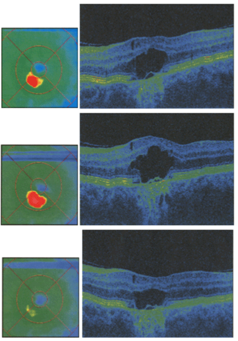
Figure 6a:
OCT scans and color-coded maps, macular edema, follow up.