|
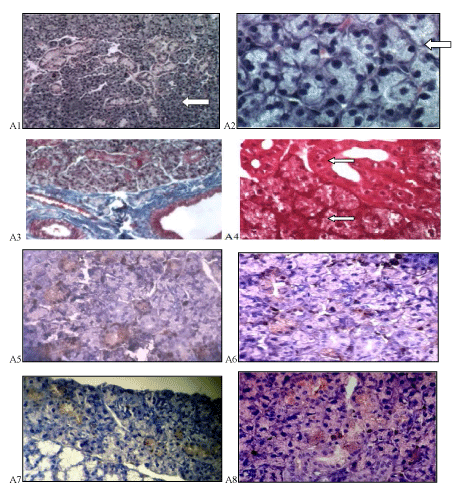
| Figure 2: Photomicrographs of rat submandibular gland of control groups showing regular structured acini (arrows) (A1, H&E ×100; A2, H&E ×400). The collagen fibers are distributed in the stroma between the acini and duct (A3, Trichrome ×100). Strong positive PAS reaction in striated duct (upper arrow) and acini (lower arrow) which is observed more at their basement membrane (A4, PAS ×400). Moderate Ki-67 immuno reactivity in nuclei of cells of ducts and some acini in the first and second group respectively (A5, A6), and moderate cytoplasmic reaction to Bcl-2 mostly seen in association with duct’s cells in the first and second group respectively (A7, A8), (immunohistochemistry ×400). |