 |
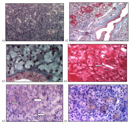
| Figure 4: A photomicrograph of a section in submandibular gland of the methotrexate and green tea extract treated group showing well formed acini and duct lining (A1, H&E ×100). The collagen fibers are distributed in the stroma between the acini and ducts, intact multiple granular convoluted tubules and intact duct lining are seen, no congested blood vessels were found (A2 Trichrome ×100, A3, Trichrome ×400). Strong positive PAS reaction in the ducts and acini with more concentration at their basement membrane, (arrow) (A4, PAS ×400). Mild Ki- 67 immuno reactivity in nuclei (arrows) of some acinar cells (A5) and moderate positive cytoplasmic reaction in duct’s cells to Bcl-2(A6), (immunohistochemistry ×400). |