 |
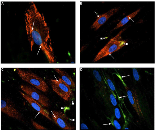
| Figure 2: Immunocytochemical localization of pro-collagen α1 type I and tenascin C proteins in HPFs treated with 0.1 mM and 0.5 mM HEMA for 15 days. CY3-conjugated anti-goat and FITCH-conjugated anti-mouse both IgG antibody were used to detect the double-localization of the proteins. All samples were counterstained with DAPI. (A) and (B) show HPFs without any treatment. Red signal correspond to CY3 pro-collagen α1 type I protein (arrow). FITC fluorescent signal (green) corresponds to tenascin-C protein; (C) Samples exposed to 0.1 mM HEMA showed pro-collagen α1 type I protein (arrow). (D) Samples treated with 0.5 mM of HEMAs showed an high signal of TNC (arrow). All the images were 600X. |