 |
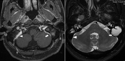
| Figure 2: T1- (left) and T2- (right) weighed axial MRI showed a well-defined, high-signal-intensity, soft-tissue mass protruding to the left posterior fossa. The left sigmoid sinus (arrow) was compressed by the cholesterol granuloma, compared with the normal right sigmoid sinus (arrowhead). |