|
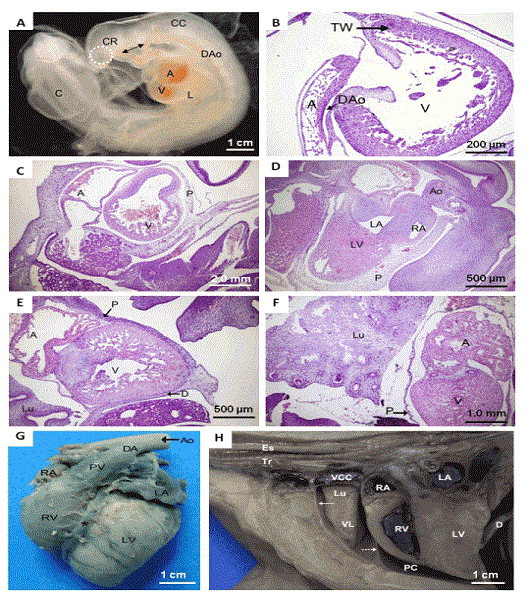
| Figure 1: (A) A 21-day embryo. Observe in the cranial region (CR) the beginning of the formation of the optical placode (circle). Note the cervical curvature (CC) and the caudal region (C). At this stage, the heart chambers are forming; the ventricle (V) and atrium (A) are still undergoing torsions. Note also the syntopy with the liver (L). Observe the extent of the dorsal aorta (DAo) and its branches (arrow) in the region of the brachial arches. (B-C) Histology of the heart after 25 days of embryo development. Note the atrium (A), ventricle (V), dorsal aorta (DAo), and trabecular wall of the ventricular chamber (TW). (D-F) Observe the chronology of the development of the embryo’s heart at 30 days, 33 days, and 38 days, respectively. Note the right atrium (RA) and left atrium (LA) and the ventricles (V). Throughout development, the muscular wall of the left ventricle (LV) greatly increases in thickness. Observe the pericardium (P), lung (Lu), aorta (Ao), and diaphragm (D). (G) Auricular face of the fetal horse heart at 105 days of gestation. Observe the right ventricle (RV), left ventricle (LV), left atrium (LA), right atrium (RA), pulmonary vein (PV), ductus arteriosus (DA), aorta (Ao), and paraconal interventricular branch of the left coronary artery (*). (H) Lateral view of the thoracic cavity of a horse fetus at 110 days of gestation. Note the fibrous pericardium with the pericardial blade of the serous pericardium (dashed arrow), pericardial cavity (PC), parietal pleura (continuous arrow), the open right atrium (RA) and right ventricle (RV), the left atrium (LA), left ventricle (LV), diaphragm (D), vena cava cranial (VCC), ventral lobe (VL) of the right lung (Lu), esophagus (Es), and trachea (Tr). |