 |
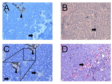
| Figure 1: A) Vasculogenic mimicry in Merkel cell carcinoma (Stage 3) indicated by the presence of intratumoral extra vascular erythrocytes (next to regular endothelial cell-lined blood vessels). Hematoxylin and eosin-stained Merkel cell tumor sections stained with CD34. Regular blood vessels are sharp by arrowheads, and areas of vasculogenic mimicry are identified by arrows (Magnification 100x). B) Vasculogenic mimicry in Merkel cell carcinoma indicated by the presence of a histological pattern detected by periodic acid Schiff (PAS)-staining (PAS loops) (magnification 100x). C) CD34 counterstained with hematoxylin and eosin Merkel cell tumor (stage 1) sections (Magnification 200x). With a zoom of the normal blood vessels (arrowheads) and bloodlakes (arrows). D) Merkel cell carcinoma (Stage 2), hematoxylin and eosin stained (magnification 100x). Bloodlake identified by arrow. |