|
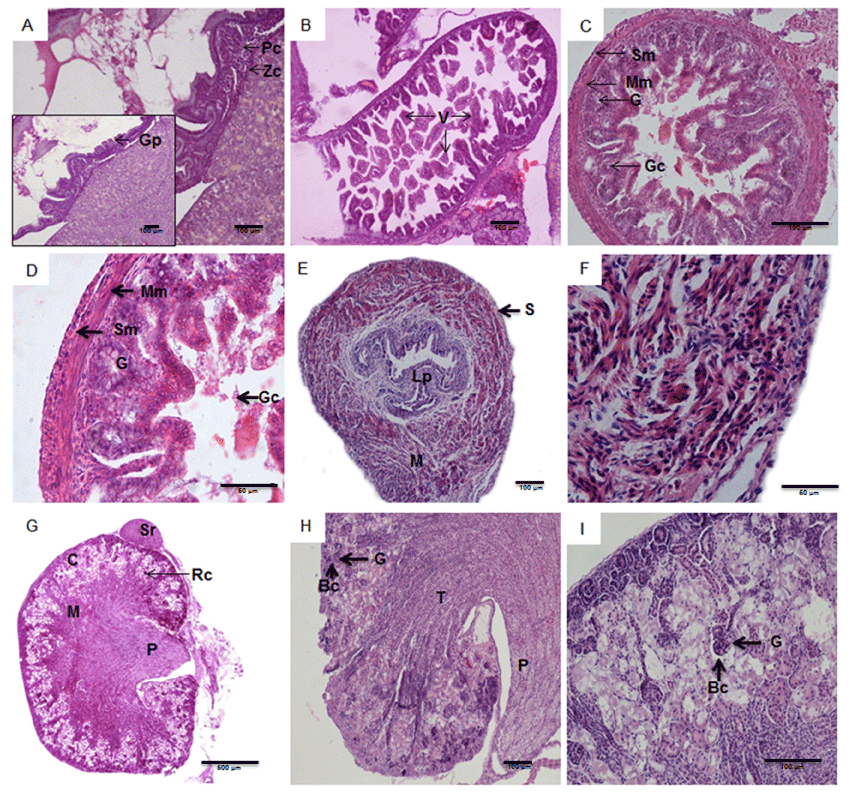
| Figure 4: A Fetus at gestational days 18 and 18.5; notice parietal cells (Pc); zymogenic cells (Zc), and gastric pit (Gp). B In the same animal, note the villi (V) that were cut in the same section as intestinal loops. C Full-term fetus gestational days 20 and 20.5; note the layers of the intestine, which consists of submucosa (Sm), muscularis mucosae (mm), gastric gland (G), and goblet cell (Gc). D, Detail of the intestine, featuring the submucosa (Sm), muscularis mucosae (Mm), gastric gland (G), and goblet cells (Gc). E Full-term fetus at gestational days 20 and 20.5; note the lamina propria (Lp) in the bladder, the muscle layer (M), and the serous layer (S). F Thick muscular layer of the bladder. G Full-term fetus at gestational days 20 and 20.5; note the kidney with the following structures: suprarenal (Sr), cortical area (C), medullary region (M), renal corpuscle (Rc), and pelvis (P). H At higher magnification, Bowman’s capsule (Bc) is observed surrounding the glomerulus (G), the longitudinal tubules (T), and pelvis (P). Detail of Bowman’s capsule (Bc), which surrounds the glomerulus (G) and makes up the renal corpuscle. |