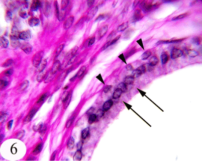
Figure 6:
A photomicrograph of the immature seminal gland showing the linning epithelium of the acini; tall columnar cells (arrow) and basal cells (arrow head). Stain: PAS Obj.x100 : Oc.x10