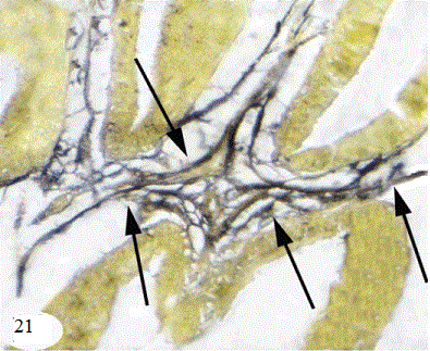
Figure 21:
A photomicrograph of the mature seminal gland showing the distribution of reticular fibers in between acini (arrow) Stain: Silver stain Obj.x40 : Oc.x10