|
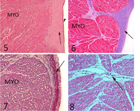
| Plate 3: Figure 5: A photomicrograph of the papillary muscle showing the endothelium (arrow head), the subendothelium (arrow) and the myocardium (MYO) Stain: H& E Obj.x5: Oc.x10. Figure 6: showing the subendothelial collagen fibers (arrow) and the myocardium (MYO). Stain: Azan Obj.x5: Oc.x10. Figure 7: showing the subendothelial elastic fibers (arrow) and the myocardium (MYO). Stain: Weigert’s Resorcin Fuchsin Obj.x10: Oc.x10. Figure 8: showing the highly vascularized inter bundle connective tissue (arrow). Stain: H&E Obj.x10: Oc.x10. |