|
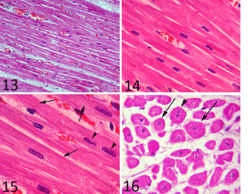
| Plate 5: Figure 13, 14 and 15: A photomicrograph of the papillary muscle myocardium showing the longitudinal section of the cardiomyocytes. Stain: 13, 14 and 15) H&E. 13) Obj.x10: Oc.x10. 14) Obj.x63: Oc.x10. Figure 15: showing the cardiac muscle fibers striation (arrow) and binucleated cells (arrow head). Obj.x100: Oc.x10. Figure 16: showing the cross section of the cardiac muscle fibers (arrow) and binucleated cells (arrow head). Stain: H& E Obj.x40: Oc.x10. |