|
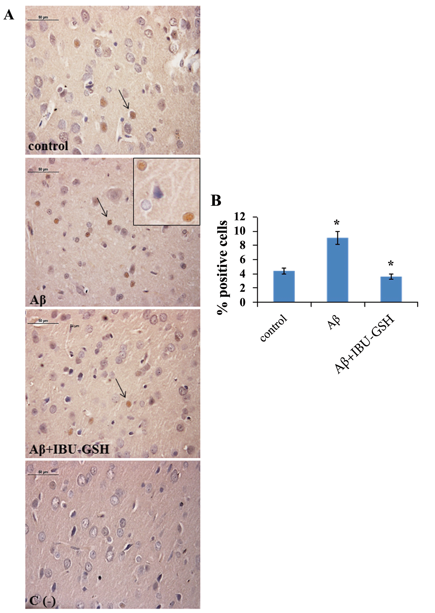
| Figure 7: A: Immunohistochemical detection of caspase-3 in rat cerebral cortex in different experimental conditions. Scale bar size 50 μm. Control: untreated sample; Aβ: Aβ-injected cerebral cortex; Aβ+IBU-GSH: Aβ-injected cerebral cortex+IBU-GSH conjugate; C (-): negative control. Insert shows nuclear localization of cleaved caspase-3 (magnification 40x); B: Graphical representation of caspase-3 positive cells percentages. Percentages values are means ± SD; * Aβ+IBU-GSH conjugate vs Aβ: p < 0.05 |