 |
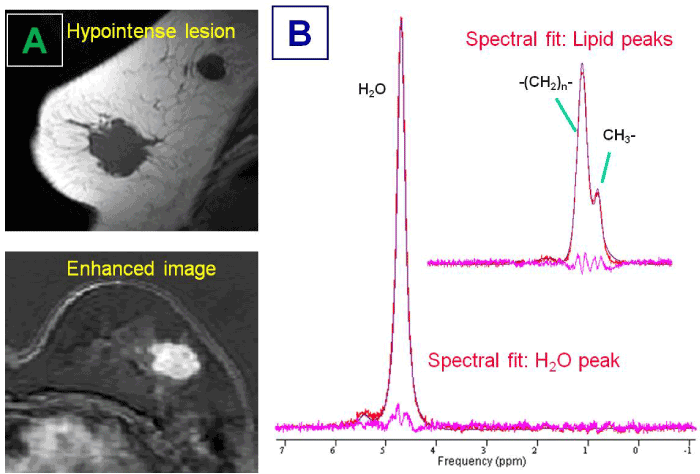
| Figure 1: Shows a representative MR imaging and 1H-MRS measurement from a patient who received chemo-follow up treatment. Free-hand core biopsy revealed an invasive ductal carcinoma. A radiologist determined the size measurement based on the maximum intensity projection (MIP) of the subtraction images. Before treatment, the lesion was 3.0 cm and showed a heterogeneous enhancing pattern (Figure 1A). Lipids peaks (e.g., methyl (-CH3) at 0.92 ppm, methylene (-CH2-) at 1.33 ppm) were clearly visible in the spectrum without water-fat suppression and fitted by using a Lorentzian model (Figure 1B). |