|
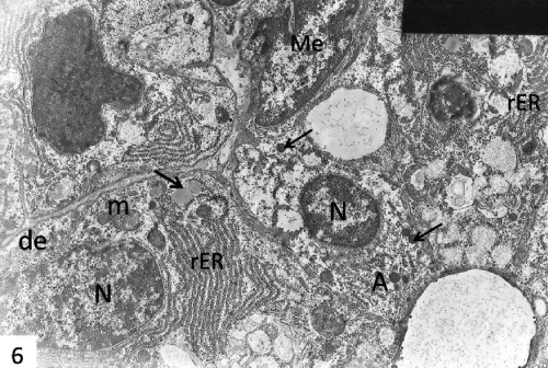
| Figure 6: An electron micrograph of control submandibular gland showing normal shaped acinar cells (A) grouped around lumens and demilunar cells (de). The cells showing vesicular nucleus (N), all the cell organoids of secretory cells, ,well developed rough endoplasmic reticulum (rER), numerous mitochondria (m), electron dense (thin arrows) and electron lucent (thick arrow) granules. Note, a myoepithelial cell (Me) surrounding the acinus. X 4000. |