 |
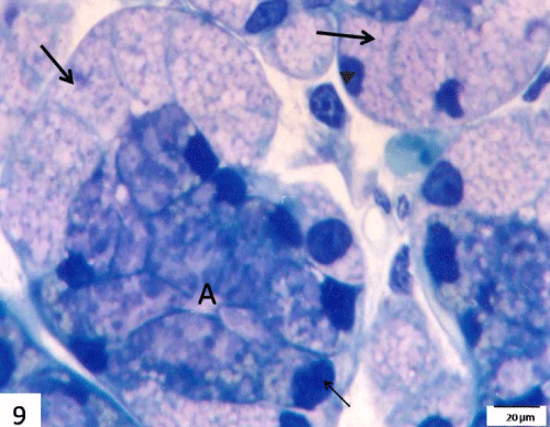
| Figure 9: A semithin section in the submandibular gland of 7 days starved group stained with toluidine blue showing acinar cells (A) with loss of metachromatic staining of its content and irregular dark stained nuclei (thin arrow). Note, markedly distended demilune cells with faintly stained metachromatic granules (thick arrows) and their small dark stained (pyknotic) nuclei (arrow head). Scale bar=20 μm. |