 |
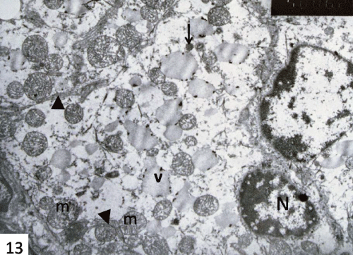
| Figure 13: An electron micrograph of the submandibular gland of 7 days starved group showing the striated duct cells with small irregular condensed nucleus (N), disrupted mitochondria (m), a few scattered fine granules (arrow) and multiple vacuoles (v). Note, the remnant of basal membrane infoldings (arrow heads). X 4000. |