 |
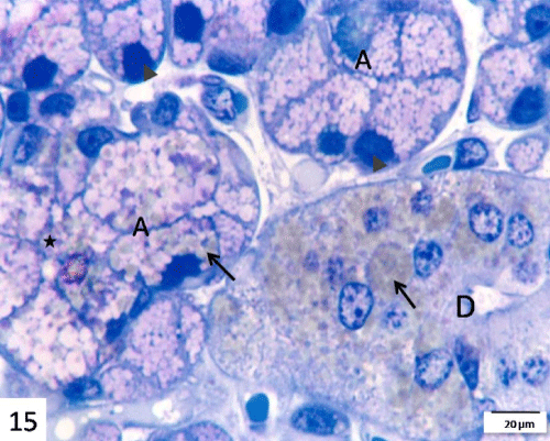
| Figure 15: A semithin section in the submandibular gland of 14 days starved group stained with toluidine blue showing irregular shaped small sized acini (A) with dark irregular nuclei (arrow heads) and lossed of both metachromatic and orthochromatic secretory material. Note, the graying lipid droplets (arrows) in acinar cells and in the cells of striated duct (D). Note, few deeply stained blue granules near the luminal surface of the acinar cells (star). Scale bar=20 μm. |