|
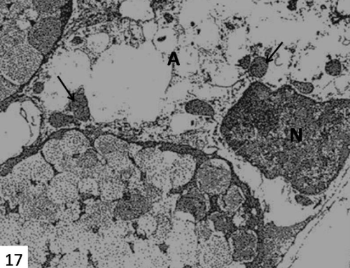
| Figure 17: An electron micrograph of submandibular gland of 14 days starved group showing sever degenerated degranulated acinar cell (A) with complete loss of all cytoplasmic organelles and irregular nucleus with amorphous appearance of its chromatin (N). Note, numerous small electron dense deposits (arrows) in the cytoplasm. X 4000. |