| Figure 3: TEM images of MSCs cultured on 3D scaffold for (A) 7 days. The
cells showed a prolonged body strictly adhering to the scaffold surface (bar:
2 μm); (B) MSCs after 14 days of 3D culture. (bar: 2 μm); (C) MSCs after 21
days of 3D culture. The cells showed several secretive vacuoles (insert) (bar:
2um; insert bar: 500 nm); (D) MSCs after 28 days of 3D culture. An elevated
deposition of extracellular matrix is shown (bar: 2 μm); (E) high magnification of
extracellular matrix in MSCs grown on a 3D scaffold for 28 days. Fibers with a
banding structure are clearly detected (bar: 200 nm); (F) TEM images of MSCs
grown on 2D culture system for 28 days. A lower deposition of extracellular
matrix is detected (bar: 1μm). High magnification of the organization of
extracellular matrix (insert; bar: 200 nm). |
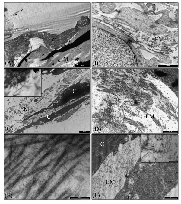 Abbreviation: (C) MSCs; (M) 3D material; (EM) extracellular matrix