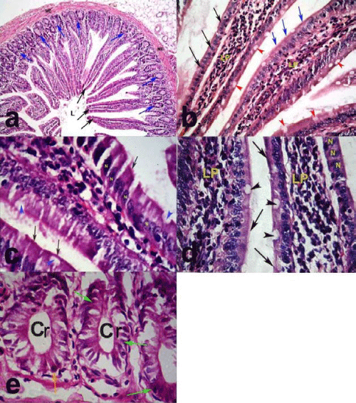
a) villi (black arrows) that appear as finger like projections thrown into the lumen
(L). The tubular crypts (blue arrows) arranged deep to the muscularis externa (ME). (H &E X 100)
b) lamina propria (LP) of the villi covered by a single layer of tall columnar cells with oval basal nuclei (black arrows). The goblet cells (arrow heads) are scattered in between the columnar cells. The brush border (blue arrows) is intact.
(H &E X 200)
c) villi lined by tall columnar cells with oval basal nuclei (N) and the goblet cells (arrow heads) are scattered in between. The brush border (arrows) is intact. (H &E X 400).
d) lamina propria (LP) of the villi covered by tall columnar cells with oval basal nuclei (N) and the goblet cells (arrow heads) are scattered in between. The brush border (arrows) is intact. (H &E X 400)
e) crypts (Cr) lined by columnar epithelial cells with basal oval nuclei (green arrows)
and Panth cell (orange arrow). (H & E X 400).