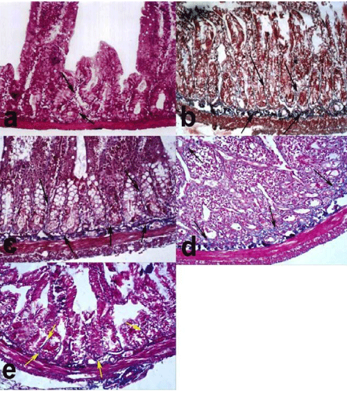
a) group I showing minimal amount of collagen fibers (arrows) in the submucosa and in between the villi.
b) group III showing increased amount of collagen fibers in the submucosa and among the crypts (arrows).
c) group IV showing increased amount of collagen fibers in the submucosa and among the crypts (arrows).
d) group V showing mild amount of collagen fibers in the submucosa (arrows).
e) group VI showing mild amount of collagen fibers among the crypts and in the submucosa (arrows). (Masson’s trichrom X 100).