 |
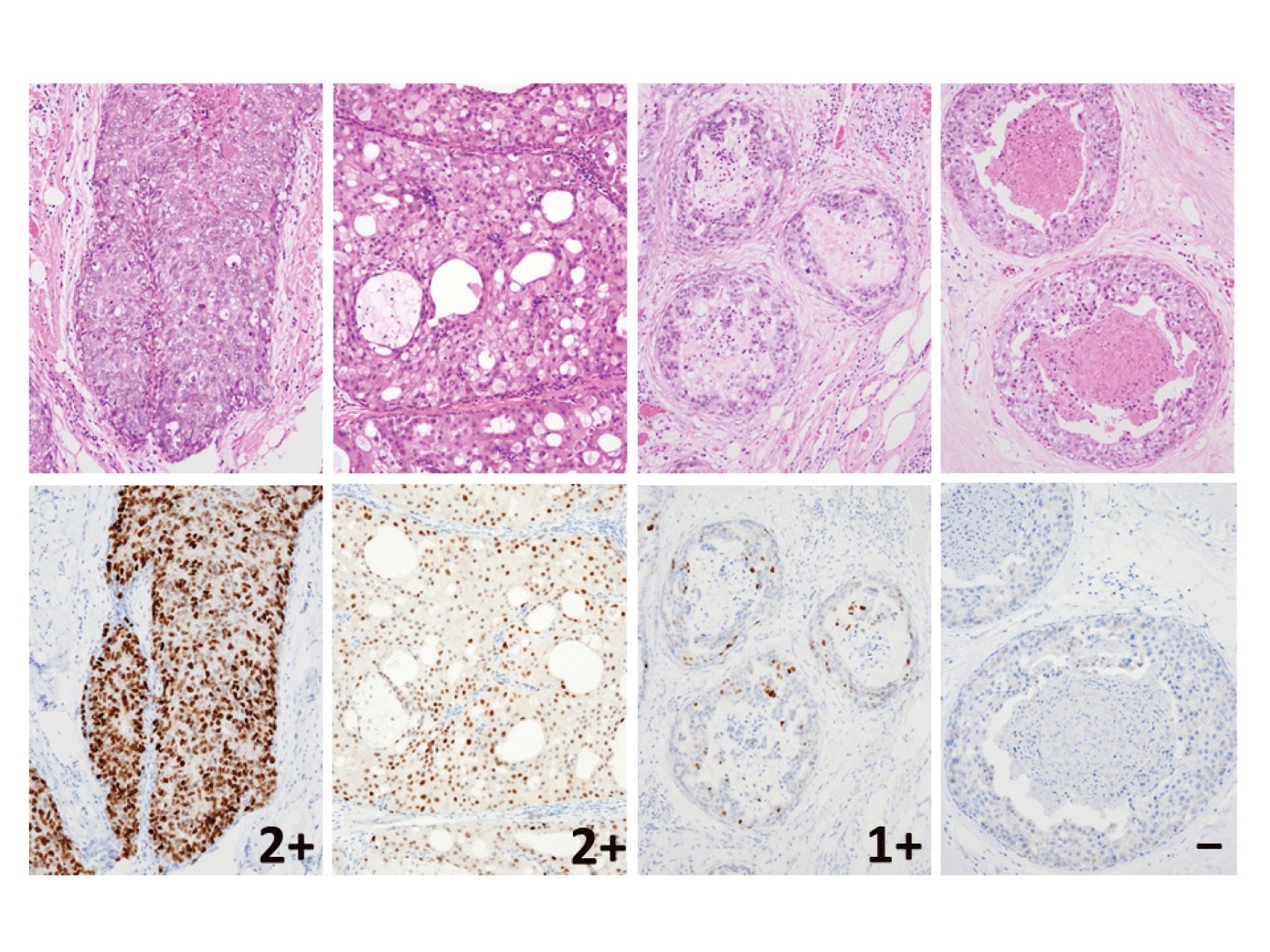
| Figure 1a: H.E.staining and P53 immunostaining pattern in DCIS. (upper) H.E.staining (lower) P53 staining corresponding to upper H.E.staining. The P53 immunostaining pattern was classified into 3 groups: 2+ (homogeneous and diffuse staining, ≥ 50% of cancer cells), 1+ (heterogeneous or focal staining, 10–49% of cancer cells), and negative (focal staining, <10% of cancer cells). |