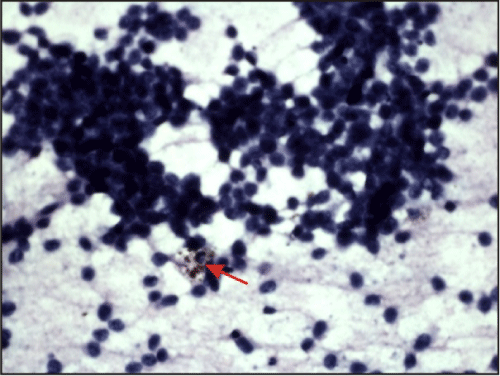
Figure 2:
[H&E 100X] smear shows malignant cells arranged in group and clusters and singly scattered. The cells have hyperchromatic pleomorphic nuclei and scanty cytoplasm. The arrow shows malignant cell containing melanin pigment.