|
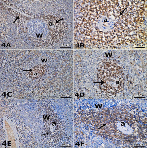
| Plate 4: Representative Micrographs of Spleen Using Immunoperoxidase Staining for CD3. (A, B) Control rat tissue showing numerous strong brown positive immunoreactions for T-lymphocytes (arrow) noticed mainly around the central artery (a) forming periarterial lymphatic sheath (PALS) in white pulp (W). (C, D) MSG-treated rat tissue showing depletion of T-lymphocytes (arrow) in PALS around central artery (a) of white pulp. (E, F) Recovery group rat tissue showing many positive T-lymphocytes (arrow) in PALS in white pulp. (Images scale bar, A,C,E; 100 μm & B, D, F; 25 μm). |