|
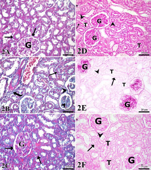
| Plate 2: A: Control group renal cortex showing thin blue collagen fibers (arrow) around the renal corpuscle (G) and tubules. B: Triclosan treated group showing excess blue-stained collagen fibers around tubules (arrow), around and within glomeruli (arrowhead). Foci of interstitial fibrosis (curved arrow) are also noticed. C: Triclosan-ellagic acid treated group showing blue collagen fibers (arrow) around the renal corpuscle and tubules. D: Control group showing intense PAS +ve reaction reaction in the basal lamina (arrow), luminal brush border (arrowheads) of the renal tubules (T) and renal glomeruli (G). E: Triclosan treated group showing strong PAS +ve reaction in the renal glomeruli (G) while renal tubules (T) show moderate reaction in basal lamina (arrow), luminal brush (arrow head) border. .F: Triclosan-ellagic acid treated group showing moderate PAS +ve reaction in the basal lamina (arrow), luminal brush (arrow head) border of the renal tubules (T) and glomeruli (G). (scale bar 25 μm, A, B, C; Masson’s trichrome stain & D, E, F; PAS stain). |