|
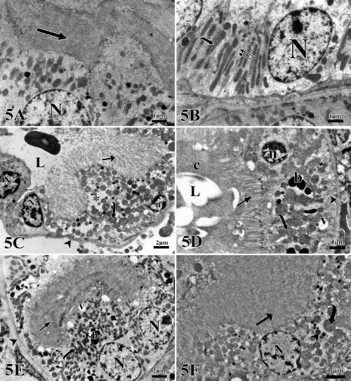
| Plate 5: Electron micrographs of a proximal convoluted tubule (PCT) cells. A, B: Control group showing a proximal convoluted tubule epithelial cells lining with large euchromatic nuclei (N), prominent nucleolus and long apical microvilli (arrow). Many elongated basal mitochondria (curved arrow) are seen in-between extensive basal infoldings (double arrows) are also noticed. All cells are rest on basal lamina. C,D: Triclosan treated group showing a disoriented basal mitochondrial (curved arrow). Multiple cytoplasmic vacuoles (v), electron-dense bodies (b) and areas of rarified cytoplasm (*) are noticed. Apically displaced nuclei with clumps of heterochromatin (N) and apoptotic nuclei (n) are detected. Casts (c) with different electron density are found at the tubular lumen (L). Apical long microvilli (arrow) are preserved on some cells, while others show sparse short microvilli (double arrows). Irregular basal lamina (arrow head) could be seen. E,F: Triclosan-ellagic acid treated group showing euchromatic nuclei (N) and mitochondria (curved arrow), electrondense bodies (b) and apical vacuoles (v). Numerous apical long microvilli (arrow) are also noticed. Regular basal lamina is also noticed (arrow head). (Images scale bar, A,B,D,F; 1 μm & C, E; 2 μm). |