 |
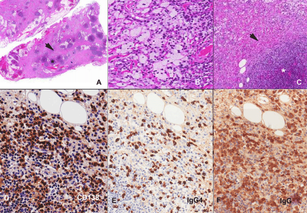
| Figure 2: (A-F): Sclerosing xanthogranulomatous inflammation form of IgG4RD. (2A): Orbital biopsy, 2x Hematoxylin & Eosin (HE) stain showing areas of fibrosis (arrow) and lymphoplasmacytic aggregates (asterix). (2B): HE stain, 40x. Xanthogranulomatous inflammation characterized by foamy histiocytes. (2C): HE stain 10x. Dense lymphoplasmacytic infiltrate (asterix) and storiform fibrosis (arrow). (2D & E): CD138 and IgG4 immunohistochemistry (Peroxidase, 20x) showing increased tissue IgG4 CD138 positive plasma cells ~102 cell/ hpf. (2E & F): IgG4 and IgG immunohistochemistry (Peroxidase, 20x) showing increased IgG4: IgG ratio (75%). |