|
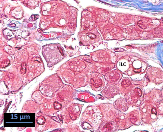
| Figure 4: Kangaroo LC from the region around the TR. Most of the cells are active cells (aLC) with foamy cytoplasm while one inactive (iLC) lipid laden cell which has two large lipid droplets, scattered blood vessels (BV) among LC. Trichrome stain, line scale=15 μm. |