|
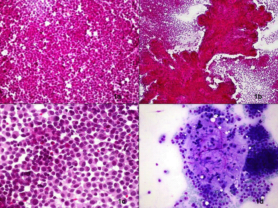
| Figure 1: a) shows stained sections neoplastic cells, b) presents the papillary structures, c) shows monomorphic cells with peripherally located inconspicuous nuclei, dispersed chromatin and the abundant granular cytoplasm, d) Mucoid material visualization. |