 |
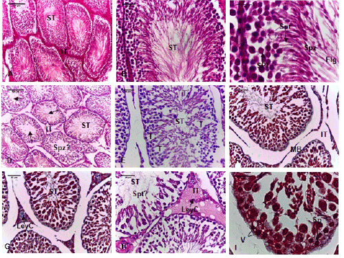
| Figure 2:Photomicrographs of rats testicular control and treated. The control (A, 100x; B, 400x and C, 1000x; H&E) reveal interstitial tissues (IT) and seminiferous tubules (ST) with normal germ cells: spermatogonia (Sp), spermatocytes I (SPI), spermatids (Spt), spermatozoa (Spz) with flagellum (Flg). The rats treated showing change in the testicular parenchyma, abundant interstitial tissues (IT) and absence of sperm in lumen of tubules (Spz?) (D, H&E 100x), empty spaces after the disappearance of sertoli cells (T) (E, H&E 400x), basal lamina is separate from the tubules and is irregular (F and G, Trichrome 400x), germ cells in seminiferous tubules and leydig cells (LeyC) decreased sharply (H, H&E 400x), vacuolization of germ cells (I, Trichrome 1000x). ST: seminiferous tubules; Sp: spermatogonia; SPI: spermatocytes I; Spt: spermatids; Spz: spermatozoa; IT: interstitial tissue; LeyC: Leydig cell; Flg: flagellum; V: vacuole; T: disappearance of sertoli cells. (A, D: Scale bar 100 µm; B, E, F, G, H: Scale bar 20 µm; I: Scale bar 10 µm) |