|
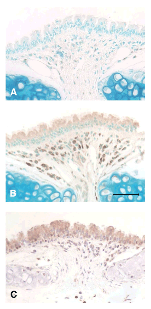
| Figure 1: Immunoperoxidase staining for IL-33 in mouse trachea. (A) Tissue from a naïve animal and (B) from an animal 4 hours after induction of an experimental acute exacerbation, both stained using goat anti-mouse IL-33 (control sections omitting the primary antibody, or replacing with normal goat serum, were completely negative) (bar=50 μm). (C) Tissue from an experimental acute exacerbation similar to B, stained using rabbit anti-human IL-33. Both antibodies show cytoplasmic staining of AEC and of inflammatory cells in the lamina propria. Original magnification × 400. |