 |
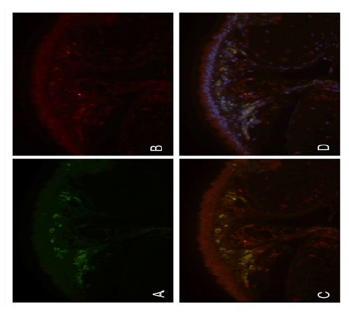
| Figure 2: Double-label immunofluorescence staining for IL-33 and IgG. (A) Inflammatory cells in the lamina propria of the trachea demonstrating immunoreactivity for cytoplasmic IgG, using an FITC-labeled antibody which fluoresces green. (B) Same field showing tracheal epithelium and inflammatory cells in the lamina propria demonstrating immunoreactivity for IL-33, using an Alexa Fluor 555-labeled antibody which fluoresces red. (C) Digitally merged image of A and B, demonstrating co-localisation of IL-33 in inflammatory cells in the lamina propria with cytoplasmic IgG, seen as yellow, confirming that these are plasma cells. Note non-colocalising staining for IL-33 in the tracheal epithelium. (D) Digitally merged image of C with a further image of the same field demonstrating staining of nuclei of epithelium and inflammatory cells, using DAPI which fluoresces blue. Original magnification ×400. |