|
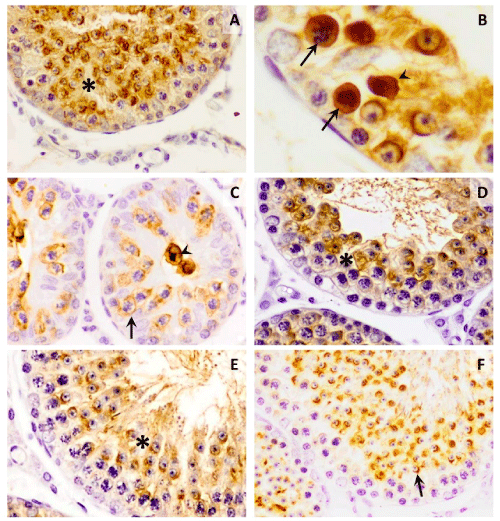
| Figure 1: Lectin histochemistry of PNA lectin with affinity by Galβ1,3-GalNAcα1 in the hamster testis. A) Section seminiferous tubule in process of regression after short photoperiod. The spermatids show higher affinity for PNA lectin (asterisk). X 20. B) With further magnification apoptotic spermatocytes (asterisk) and spermatids (double asterisk) are identified. X60. C) Sections of seminiferous tubules completely regressed. The affinity for the PNA lectin is shown in the plasma membrane of some spermatocytes (arrow) and some round spermatids (double arrow.X20 D) Section seminiferous tubule in process of initial recrudescence after of regression due short photoperiod. The epithelium in this portion of tubule is still in spermatids arrest. The spermatids are highly positives by PNA lectin (asterisk).X40. D) However, other section in recrudescence show portion of tubule with a decrease of both spermatids and spermatocyte in affinity for PNA (asterisk).X20. E) Final recrudescence. The lectin PNA shows affinity in spermatids, especially in its acrosome (arrow).X20. |