|
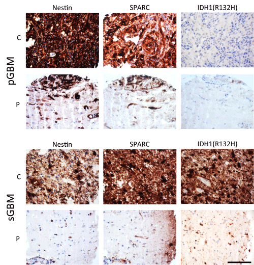
| Figure 7: Representative micrographs of tumors in pGBM and sGBM immunostained for nestin, SPARC and IDH1(R132H). The case of sGBM was positive for all three markers, whereas pGBM was IDH1(R132H) negative. The center (c) and periphery (p) of the tumors are shown. Original magnification x400. Scale bar: 100 μm. |