|
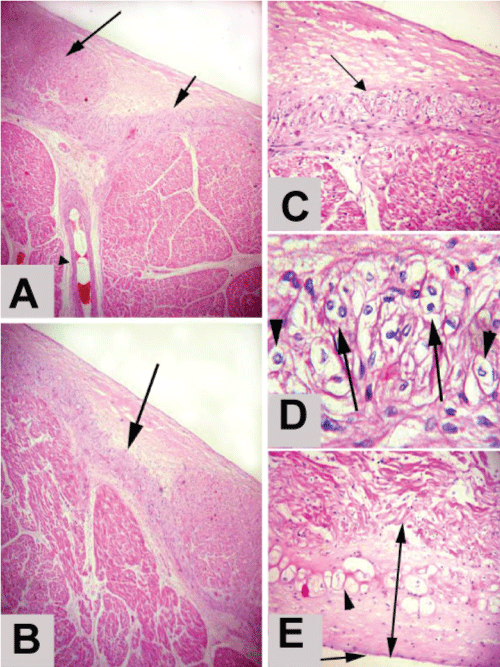
| Figure 6: (A) A photomicrograph of the terminal crest epicardium showing the sinu-atrial node head (long arrow), arm (short arrow) and BVs (arrow head) Stain: H& E Obj. x4: Oc.x10. (B) showing the long and extended arm (arrow) Stain: H& E Obj.x4 : Oc.x10. (C) showing the SAN arm cells; atrial purkinje like cells (arrow) Stain: H& E Obj.x10 : Oc.x10. (D) High magnification of (Fig. 21) showing the atrial purkinje like cells; uninucleated (arrow heads) and binucleated (arrows) Stain: H& E Obj.x40: Oc.x10. (E) A photomicrograph of the terminal crest epicardium showing the subepicardium (double head arrow), atrial purkinje like cell (arrow head) and the mesothelium (arrow) Stain: H& E Obj.x10:Oc.x10. |