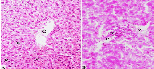
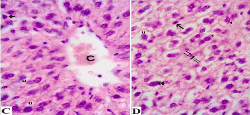
C and D Photomicrographs of ethanol-treated liver sections showed degenerating hepatocytes with fatty changes vacuoles (u) and eosinophilic alcoholic hyaline Mallory bodies (arrowheads) which appeared condensed with basophilic disintegrating cytoplasmic content (double arrowheads) in some other cells. Hepatic blood sinusoids were not apparent between the vacuolated hepatocytes and the disfigured architecture. Apoptotic cells were shrunken and numerous (arrows) with pyknosis and karyorrhexis of nuclei (double arrows). (1000X, oil immersion, Hx&E).