|
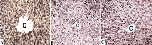
| Figure 5: Photomicrographs of Iron-Hx. stained liver sections showed A: control group with normal intracellular mitochondrial content and distribution, B: ethanol-treated rats showed moderate depletion of mitochondrial content, and C: zinc/ethanol-treated rats explored mild mitochondrial affection. Higher mitochondrial affinity was apparent in zone 2 (transparent arrows) than that in the centrilobular zone 3 (dark arrows) around the central vein (C) in the three animal groups. Blood sinusoids (S) were not apparent in ethanol-treated rats. (X400, Iron-Hx). |