|
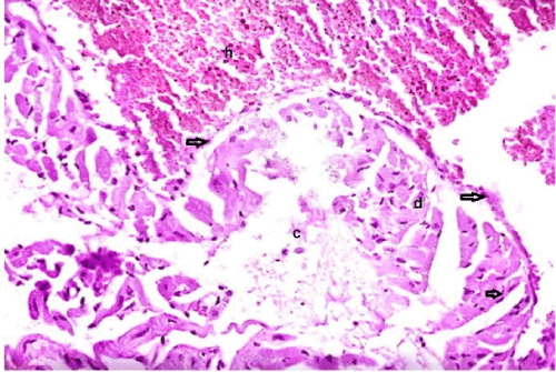
| Figure 4: A photomicrograph of a section in the cardiac muscle from group II showing wide separated cardiac myocytes, most of which appear degenerated (c), edema with pale acidophilic sarcoplasm and peripheral small dark nuclei (arrows). Notice presence of focal haemorrhage (H&E 200X). |